
Electron and Optical Microscopy
KeyLab coordinators:
Prof. Dr. Hans-Werner Schmidt
Scanning electron microscopy (SEM)
Phone: +49 (0)921 / 55-3200
E-mail: hans-werner.schmidt@uni-bayreuth.de
Homepage: Chair of Macromolecular Chemistry I
Prof. Dr. Josef Breu
Transmission electron microscopy (TEM)
Phone: +49 (0)921 / 55-2530
E-mail: josef.breu@uni-bayreuth.de
Homepage: Chair of Inorganic Chemistry I
Prof. Dr. Jürgen Köhler
Optical microscopy (OM)
Phone: +49 (0)921 / 55-4000
E-mail: juergen.koehler@uni-bayreuth.de
Homepage: Spectroscopy of soft matter
KeyLab scientist:
Dr. Ulrich Mansfeld (SEM, TEM)
Phone: +49 (0)921 / 55-4432
E-mail: ulrich.mansfeld@uni-bayreuth.de
Dr. Markus Drechsler (TEM)
Phone: +49 (0)921 / 55-3188
E-mail: markus.drechsler@uni-bayreuth.de
Dr. Lisa Günther (OM)
Phone: +49 (921) / 55-4040
E-mail: lisa.guenther@uni-bayreuth.de
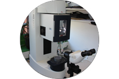
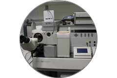
Optical microscopy (OM)
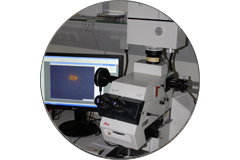
Fluorescence and Fluorescence Lifetime Imaging (FLIM) Microscope PicoQuant MicroTime 200
- Optical core: Olympus inverted research microscope with 4 objectives
- Confocal unit and piezo stage for z stacks
- Excitation sources: 4 diode lasers at 405, 485, 561, and 640 nm (both pulsed and cw operation possible)
- Two fast detectors: a single-photon counting APD module (SPAD) and a fast PMA hybrid detector with 45% quantum efficiency for time-resolved measurements
- Fluorescence spectrometer with thermoelectrically cooled EMCCD camera
- Single-molecule sensitivity
- Software for uorescence lifetime imaging (FLIM), FLIM-FRET, and uorescence correlation spectroscopy (FCS)
Standard Microscope Olympus BX 60
Optical microscope for standard applications; polarized epi- and trans-illumination
Correlation Microscope Zeiss Axio Imager A2m
Precise determination of the sample coordinates for detailed study in an electron microscope Zeiss Ultra Plus 55
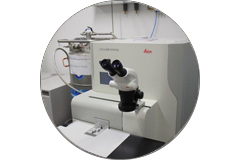
Fluorescence Microscope Leica DMR
Versatile fluorescence microscope with several spectral ranges of excitation and detection; spectrometer unit
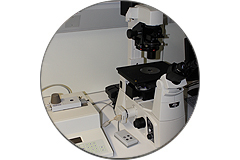
Inverted Polarization Microscope Nikon Eclipse Ti-S
Inverted polarization microscope with epi- and trans-illumination; Amici-Bertrand lens for conoscopic studies of crystals
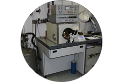
Transmission electron microscopy (TEM)
Microscopy
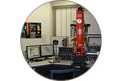
Zeiss CEM902 (TEM I)
Energy filtering transmission electron microscope (EFTEM), high voltage 50kV or 80kV, thermal tungsten cathode, incolumn Henry-Castaing energy filter, CCD camera system (Gatan Orius) with Digital Micrograph Software.
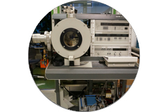
Zeiss / LEO EM922 Omega (TEM II)
Energy filtering transmission electron microscope (EFTEM), high voltage 120kV, 160kV, 200kV, thermal LaB6 cathode, incolumn magnetic energy filter of Omega type, CCD camera system (Gatan Ultrascan 1000) with Digital Micrograph Software
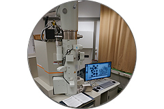
JEOL JEM-2200FS (TEM III)
Energy filtering transmission electron microscope (EFTEM), high voltage 160kV, 200kV, Schottky field emission cathode, incolumn magnetic energy filter of Omega type, CMOS camera system (Gatan OneView) with Digital Micrograph Software
Preparation
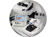
Leica Ultramikrotom UC7+FC7
Ultramicrotome for ultrathin sectioning of soft matter samples by glass or diamond knives. In an attachable cryo chamber cryo fixed liquid or semi solid samples can be sectioned at low temperatures.
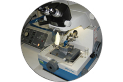
Reichert-Jung Ultramicrotome E+FC4
Ultramicrotome for ultrathin sectioning of soft matter samples by glass or diamond knives. In an attachable cryo chamber cryo fixed liquid or semi solid samples can be sectioned at low temperatures.
Jeol Cryo Ion Slicer
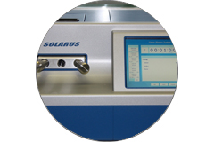
Gatan Solarus Model 950
Plasma Cleaner to remove adsorbed carbonhydrates on sample and TEM holder surfaces, hydrophilisation of surfaces.
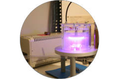
Plasma Glow Discharge unit (homemade – Biozentrum Basel)
Plasma Cleaner to remove adsorbed carbonhydrates on sample and TEM holder surfaces, hydrophilisation of surfaces.
Bal-Tec Propanjet JFD030
Cryo fixation device for liquid or semi-solid aqueous samples by a low temperature liquid propane jet.
Cryo fixation device (homemade on the base of the Zeiss Cryobox)
Cryo fixation device for aqueous liquid or semi-solid samples by plunging into liquid ethane and for samples in organic solvents in liquid nitrogen. An environmental chamber under controlled temperature and humidity conditions can be used for special samples.
Leica Kryofixierungsanlage EMGP
Cryo fixation device for aqueous liquid or semi-solid samples by plunging into liquid ethane. An environmental chamber under controlled temperature and humidity conditions can be used for special samples.
Bal-Tec Freeze Fracture device BAF400T
Metallizer for evaporation of Pt or Pt/Ir heavy metal films on inner surfaces after freeze fracture of cryo fixed liquid or semi solid aqueous samples (replica).
Bal-Tec Freeze Fracture device BAF060
Metallizer for evaporation of Pt or Pt/Ir heavy metal films on inner surfaces after freeze fracture of cryo fixed liquid or semi solid aqueous samples (replica).
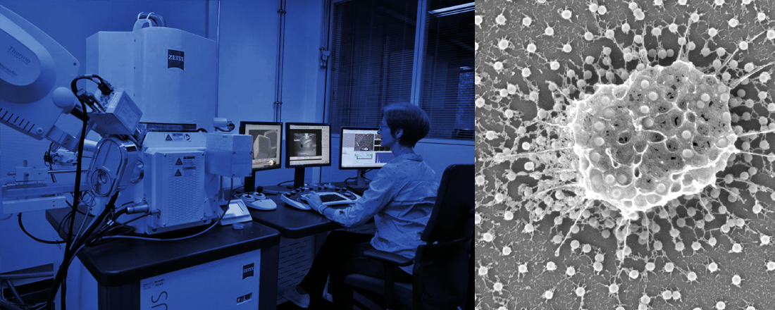
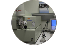
Scanning electron microscopy (SEM)
Microscopy
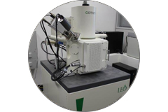
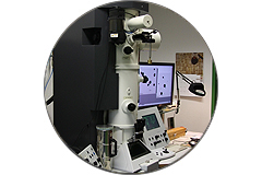
Zeiss Leo 1530 (SEM I)
High resolution FEG (field emission gun) scanning electron microscope with inlens SE-detector, chamber SE-detector (Everhart-Thornley), BSE-detector (Centaurus), minicathodoluminiscence detector and UltraDry EDS-detector (Thermo Fisher Scientific, 60 mm²)
Equipped with electron beam lithography and correlative microscopy (Zeiss Shuttle & Find).
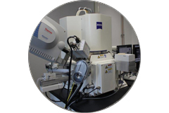
Zeiss Ultra plus (SEM II)
High resolution FEG (field emission gun) scanning electron microscope with 80 mm air lock, inlens SE-detector, chamber SE-detector (Everhart-Thornley), inlens EsB-detector (energy selective backscattered electrons), AsB-detector (angle selective backscattered electrons, 4 quadrants) and STEM-detector (Scanning Transmission Electron Microscopy). There is a gas injection charge compensation system for charging samples as well as a Peltier cooled UltraDry EDS-detector (30 mm²) and a MagnaRay WDS-spectrometer (both Thermo Fisher Scientific) for elemental analysis.
Equipped with a Leica cryo system (stage and transfer unit VCT 100) and correlative microscopy (Zeiss Shuttle & Find).
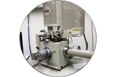
FEI Quanta FEG 250 (SEM III)
High resolution FEG (field emission gun) scanning electron microscope with ETD (Everhart-Thornley), CBS detector (concentric backscattered detector), ICD (In-column SE detector) and STEM-detector for high vacuum, as well as a LFD (Large Field detector) and CBS for low vacuum.
For ESEM mode (environmental electron microscopy) the microscope has a Peltier cooling stage and a GSED (gaseous secondary electron detector), an ESEM GAD (gaseous analytical detector for backscatter and secondary electrons) and a WET-STEM detector.
Equipped with an UltraDry EDS-detector (Thermo Fisher Scientific, 100 mm²).
Preparation
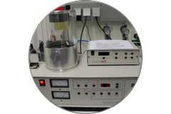
Cressington Platinum-Sputter Coater 208HR with planetary stage and quartz crystal for thickness measurement
Leica EM ACE 600 coater with planetary stage and quartz crystal for thickness measurement
Can be used either for carbon coating (carbon threads) or sputter coating (platinum) or sequential.
Leica MED 020 Preparation freeze fracture and sputter coater (tungsten) system
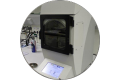
Leica EM HPM 100 High pressure freezer

Leica EM TXP Target Surfacing System
The KeyLab Electron and Optical Microscopy combines the scientific expertise of a variety of modern microscopy techniques. High resolution scanning (coordinator: Prof. Hans-Werner Schmidt) and transmission electron microscopes (coordinator: Prof. Josef Breu) including cryo techniques are available for structural and morphological analysis.
The KeyLab Electron and Optical Microscopy comprises also various optical microscopes (coordinator: Prof. Jürgen Köhler) for standard applications, spatially resolved fluorescence and Raman spectroscopy, as well as correlative microscopy. Advanced methods for the preparation of liquid, gel, and polymer samples are available.
KeyLab coordinators:
Prof. Dr. Hans-Werner Schmidt (SEM), Prof. Dr. Josef Breu (TEM), Prof. Dr. Jürgen Köhler (OM)
KeyLab scientists:
Dr. Ulrich Mansfeld (SEM, TEM)
- CV and Publications
Dr. Markus Drechsler (TEM)
Dr. Lisa Günther (OM)
KeyLab-Flyer:
Rules of use:
Members of the University of Bayreuth can find the current rules of use with user fees, the application for use, and further information on booking and managing measurement appointments via the intranet
Non-affiliates of the University of Bayreuth can obtain the rules of use from the relevant KeyLab coordinators or KeyLab researchers.